News archive

Felix's paper on Structural Basis of the Pancreatitis-Associated Autoproteolytic Failsafe Mechanism in Human Anionic Trypsin is published
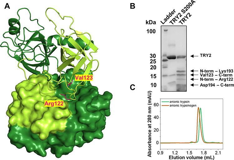
Figure 1 Structure of the TRY2 homodimer. (A) TRY2 N-terminus-Arg122 is colored yellow-green and Val123-C-terminus is colored in dark-green. The TRY2 – TRY2 complex shows one monomer in cartoon representation and one in surface representation. One monomer binds on top of TRY2 in a substrate-like manner and is rotated approximately 180° around the y-axis. Arg122 and Val123 are highlighted in pink. (B) SDS-PAGE of activated TRY2 WT and the catalytically inactive S200A variant. (C) Analytical size exclusion chromatograms of anionic trypsinogen and activated anionic trypsin.
Figure 2 Arg122-Val123 cleavage site and specific interactions of the TRY2 dimer interface. (A) Close-up of the cleaved Arg122 loop. The 2Fo-Fc density map is shown 1.6 Å around Arg122 and Val123 and is contoured at 1.0 σ. No additional density for an intact loop is visible. (B) Interaction interface with interacting residues represented as sticks. The color scheme is the same as in Figure 1. (C) Arg122 specifically interacts with Asp194 in the S1 binding pocket. The 2Fo-Fc density is shown using the same parameters as in (A). (D) Crystallographic assembly of a TRY2 fiber with repetitive TRY2 dimers. Arg122 is highlighted in red. Two nicked TRY2 monomers (light and dark grey) form the asymmetric unit.

Felix's paper on Structural Basis of the Pancreatitis-Associated Autoproteolytic Failsafe Mechanism in Human Anionic Trypsin is published in Journal of Inflammation Research. Congratulations Felix!

fulltext


Back

aktualisiert am 10.10.2024 durch
N. Geist