
International collaboration uncovers molecular “zipper” controlling a key blood-clotting enzyme

Graphical Abstract
Opening-/Closing animation
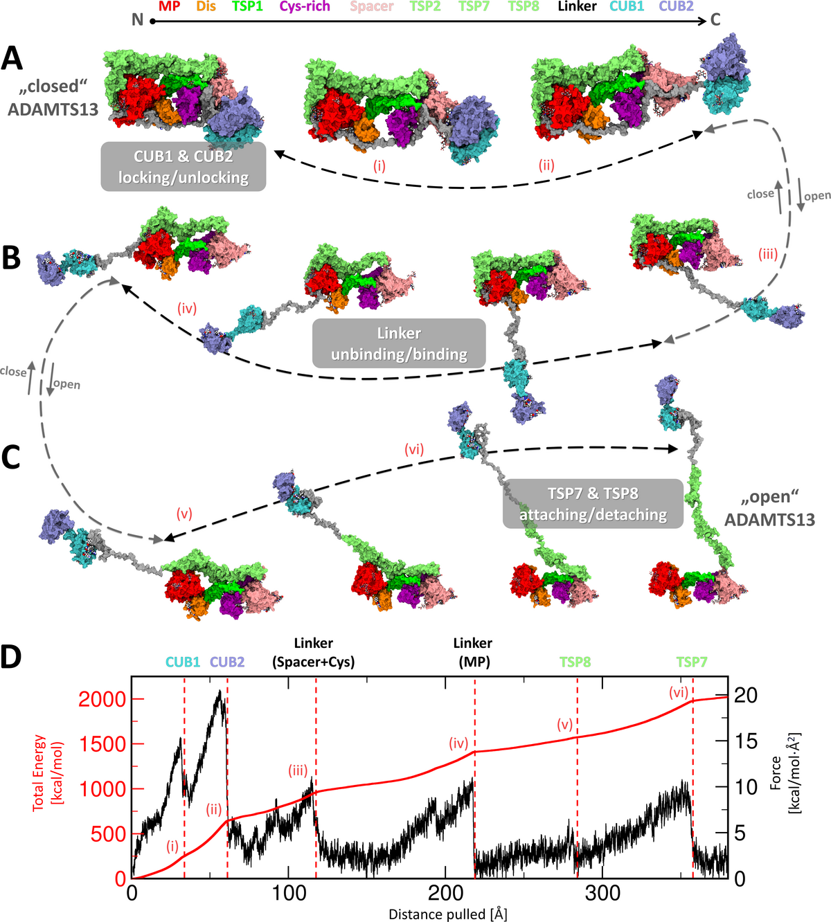
Figure 6: Conformational activation and inactivation mechanisms during global latency of ADAMTS13. Domain colors and sequential order are indicated at the top. (A) (left) Our final closed model of ADAMTS13-Del3To6 shows both terminal CUB domains attached to Spacer, the Linker region covers the substrate-binding sites on the MP, Dis, and Cys-rich domains, while TSP7 and TSP8 bound near the MP domain. Pathway for conformational activation/inactivation illustrated with arrows. A movie illustrating this process intuitively is included in the provided Zenodo archive. During activation, the CUB1 and CUB2 domains detach from the Spacer module. Roman numerals (i-vi) highlight major rupture events observed during forced unfolding simulation (Sim6 ). (B) The Linker region further disengages, exposing substrate-binding exosites on the Cys-rich, Dis, and MP domains. (C) The TSP8 and TSP7 domains detach from the MP domain, completing the conformational activation of ADAMTS13. (D) Exemplary force and accumulated energy profiles derived from the forced unfolding simulation. (Structural snapshots relaxed with MD for natural appearance)
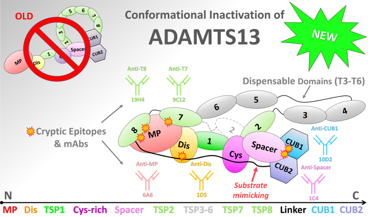
Figure 7: Conformation testing of full-length ADAMTS13 under environmental perturbations using ELISA. (top) Schematic depiction of updated model on conformationally latent ADAMTS13. Domain colors and sequential order are indicated at the top. The CUB domains rest at Spacer. Other distal domains are wrapped around the substrate binding domains and shield off various cryptic epitopes for which mAbs were recently identified. The TSP3-6 repeats are dispensable. The Linker is a pseudosubstrate, acting like VWF-A2, binding to substrate exosites of MP, Dis, and Cys-rich modules. TSP7 and TSP8 rest at the MP domain. (A) Closed plasma ADAMTS13 incubated at pH 7.2 (filled bars) or pH 6.0 (open bars). In ELISA, binding of mAbs 6A6, 1D5, 1C4, 9C12, 19H4 and 10D2, directed against the MP, Dis, Spacer, TSP7, TSP8 and CUB1 domains, was assessed. All mAbs potently bind to open, but not to closed ADAMTS13. (B) Closed plasma ADAMTS13 was incubated in the absence (filled bars) or presence (open bars) of EDTA. In ELISA, binding of mAbs was assessed as previously. Chelation of Zn2+ and Ca2+ ions destabilized the structure of the MP domain resulting in abolished binding for mAbs 3H9 and 6A6, leading to partial unwrapping of the expected domains. (relative binding (mean ± one standard deviation) to open ADAMTS13 mediated by mAb 18H10. Significance tested using two-way ANOVA, including multiple comparisons with Bonferroni correction. All experiments were performed in triplicates (n=3))

We have uncovered a previously enigmatic long-range regulation mechanism of ADAMTS13, a key enzyme that prevents dangerous blood clot formation in the circulation. Our study, titled “Wrapping it Up: Structural Basis of ADAMTS13 Global Latency”, now published in the Journal of Thrombosis and Haemostasis, provides the most detailed structural model to date explaining how ADAMTS13 maintains its inactive “closed” state.

Using large-scale molecular dynamics simulations together with biochemical experiments, we discovered that ADAMTS13 is controlled through a coordinated network of interactions across multiple domains of the enzyme. Central to the mechanism is the so-called “L3 Linker,” which acts as a pseudosubstrate and blocks critical substrate-binding regions. We describe this architecture as a “molecular zipper” that stabilizes the enzyme in its inactive state while still allowing rapid activation when needed.

The computational work was enabled by TIGER2hPE, a custom-developed enhanced-sampling simulation algorithm developed at the University of Greifswald. Even with this approach, the project required several cumulative years of computation time on the Göttingen high-performance computing infrastructure, part of NHR-NORD.

The study was coordinated by Norman Geist under the supervision of Mihaela Delcea in collaboration with Quintijn Bonnez and Karen Vanhoorelbeke at KU Leuven.

Beyond advancing the fundamental understanding of blood coagulation, our findings may support future precision therapies for thrombotic diseases such as thrombotic thrombocytopenic purpura (TTP).

Fulltext available at https://www.sciencedirect.com/science/article/pii/S1538783626003272


Zurück zu allen Meldungen