Felix's paper on Identification of Kazal Inhibitor Scaffolds with Identical Canonical Binding Loops and Their Effects on Binding Properties is published
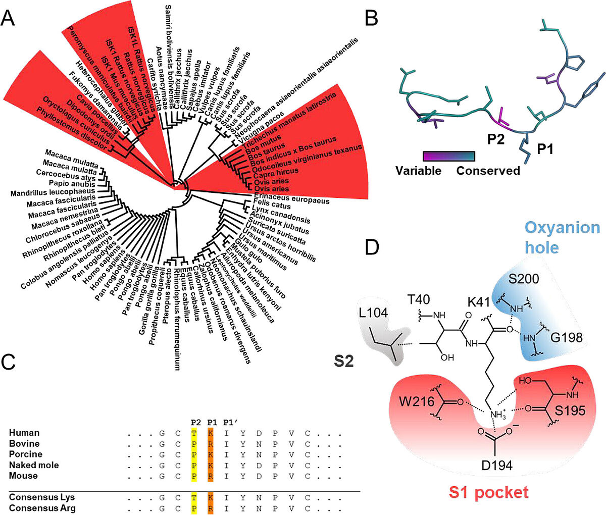
Figure 1. SPINK1 sequence and structure conservation. (A) Phylogenetic analysis of trypsin-targeting SPINK1 homologues. Duplicates indicate different isoforms within the same species. SPINK1 variants featuring arginine as the P1 residue are highlighted in red. (B) Structure of the canonical binding loop (PDB ID: 7QE8) colored according to conservation using the multiple sequence alignment from (A). Conservation scores were determined using the ConSurf server. (28) (C) Multiple sequence alignment of selected SPINK1 variants and consensus sequences of (A) subclustered according to P1 residues. (D) Structure of binding pockets in trypsin. The S1 pocket is highlighted in red, the oxyanion hole in blue, and L104 interacting with the P2 residue in gray.
Inhibitor–protease pairs form one highly integrated unit, and a secondary scaffold screen after optimizing the canonical binding loop is advisable. Optimizing the binding loop supporting scaffold can increase affinity, specificity, pH resistance, and complex stability, resulting in higher potency inhibitors. This also helps transferring results obtained from animal models to clinical trials...
Felix's paper on Identification of Kazal Inhibitor Scaffolds with Identical Canonical Binding Loops and Their Effects on Binding Properties is published in Biochemistry. Congratulations Felix!