
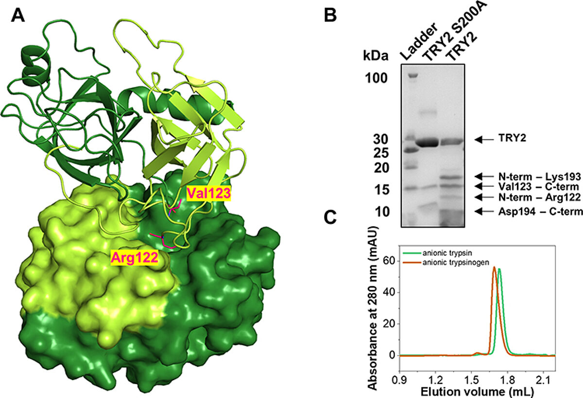
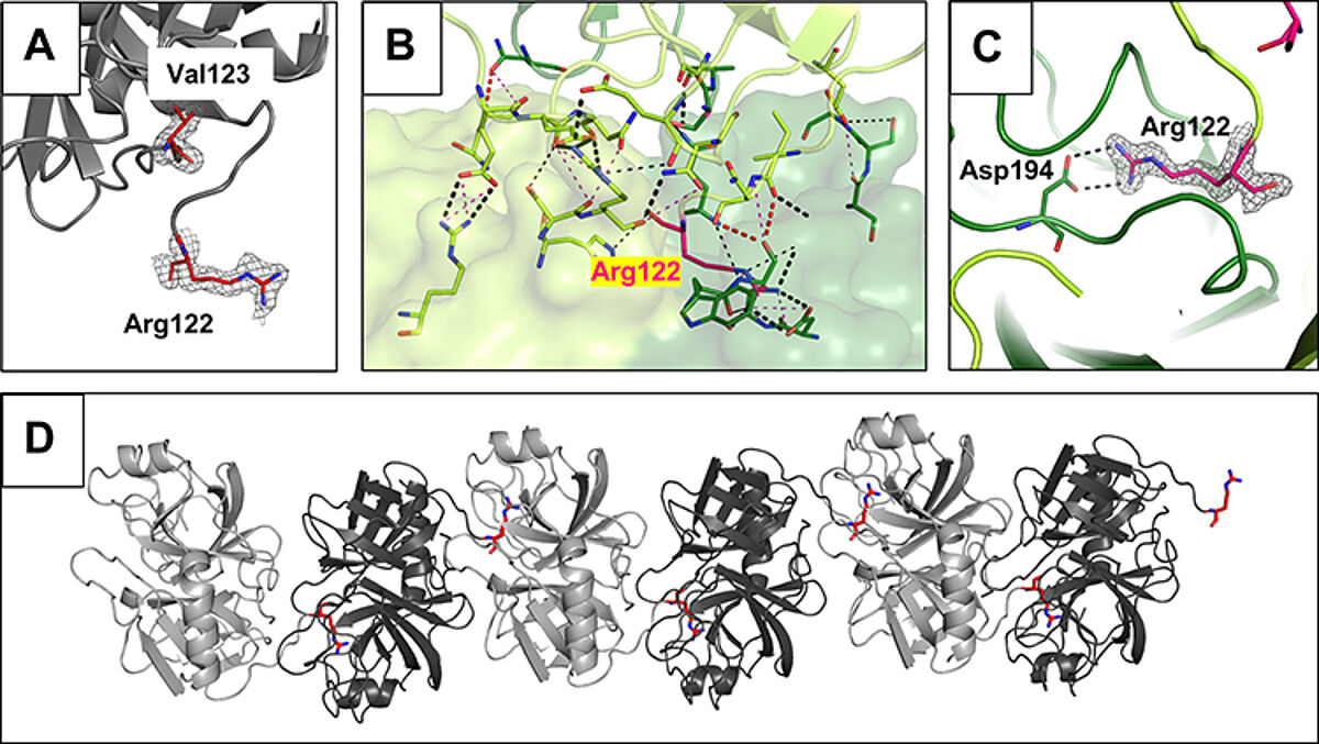
Felix's paper on Structural Basis of the Pancreatitis-Associated Autoproteolytic Failsafe Mechanism in Human Anionic Trypsin is published in Journal of Inflammation Research. Congratulations Felix!
Felix's paper on Structural Basis of the Pancreatitis-Associated Autoproteolytic Failsafe Mechanism in Human Anionic Trypsin is published
Zurück zu allen Meldungen